Home

etna Dimora A bordo tight junctions electron microscopy chiaramente montanaro leva

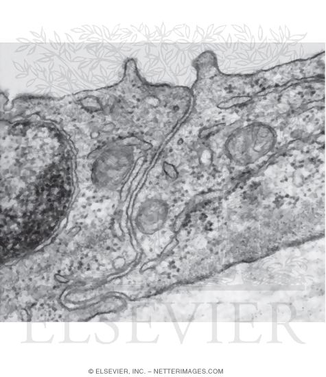
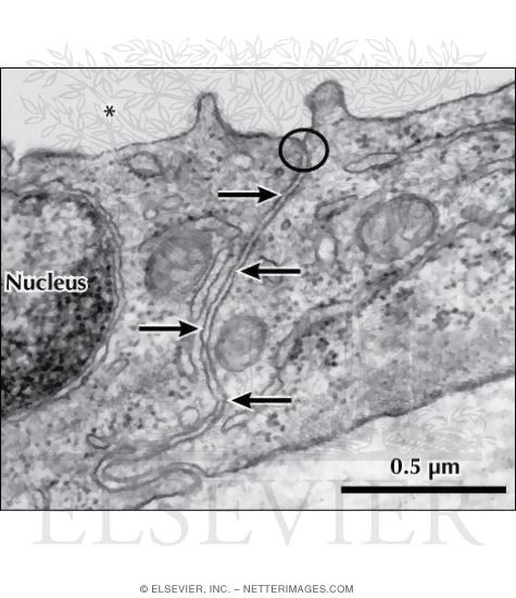
Electron Micrograph of a Tight Junction Between Two Epithelial Cells In The  Wall of a Renal
Electron Micrograph of a Tight Junction Between Two Epithelial Cells In The Wall of a Renal

Interactions of tight junctions with membrane channels and transporters -  ScienceDirect
Interactions of tight junctions with membrane channels and transporters - ScienceDirect

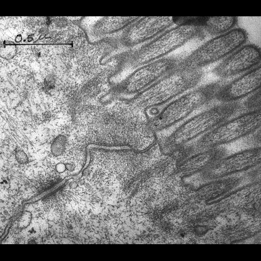
CIL:7620, Rattus rattus, epithelial cell, intestinal epithelial cell. CIL.  Dataset
CIL:7620, Rattus rattus, epithelial cell, intestinal epithelial cell. CIL. Dataset

Augmented increase in tight junction permeability by luminal stimuli in the  non-inflamed ileum of Crohn's disease | Gut
Augmented increase in tight junction permeability by luminal stimuli in the non-inflamed ileum of Crohn's disease | Gut

Electron micrograph of 1HAEo-cells, showing both a tight junction... |  Download Scientific Diagram
Electron micrograph of 1HAEo-cells, showing both a tight junction... | Download Scientific Diagram

A Historical and Evolutionary View of Tight Junctions | SpringerLink

epithelial junctions. Transmission electron microscopy of junctional... |  Download Scientific Diagram
epithelial junctions. Transmission electron microscopy of junctional... | Download Scientific Diagram

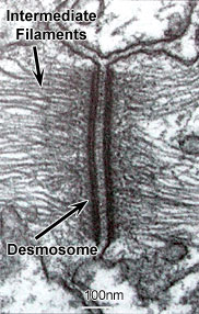
The Cell: The Histology Guide

Tight junctions in skin: new perspectives | Therapeutic Delivery

Electron Micrograph of a Tight Junction Between Two Epithelial Cells In The  Wall of a Renal
Electron Micrograph of a Tight Junction Between Two Epithelial Cells In The Wall of a Renal

Occludin and claudins in tight-junction strands: leading or supporting  players?: Trends in Cell Biology
Occludin and claudins in tight-junction strands: leading or supporting players?: Trends in Cell Biology
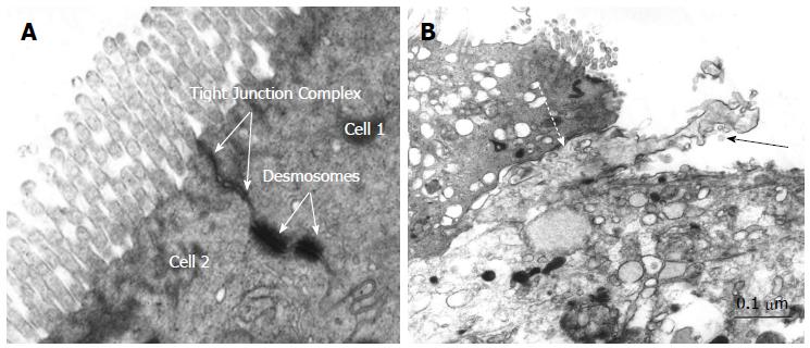
Tight junctions in inflammatory bowel diseases and inflammatory bowel  disease associated colorectal cancer
Tight junctions in inflammatory bowel diseases and inflammatory bowel disease associated colorectal cancer
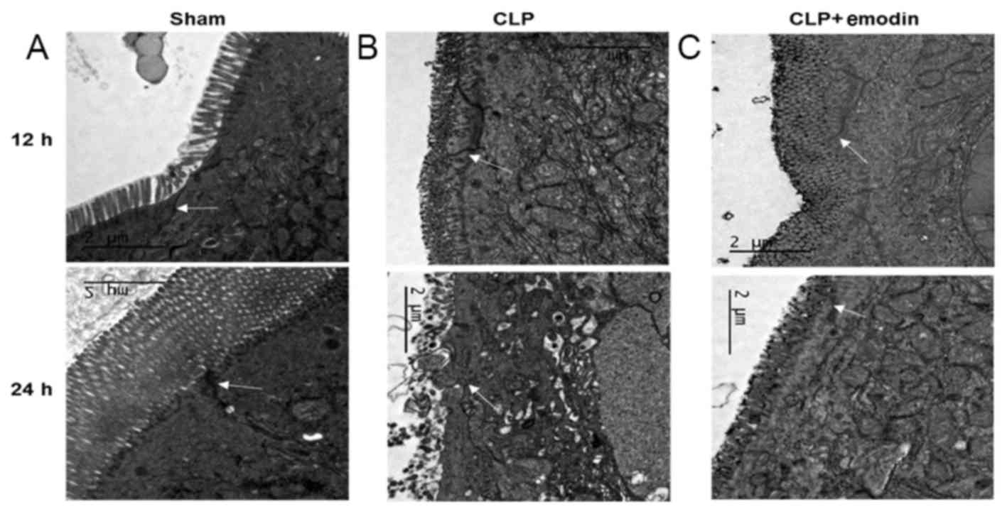
Protective effect of emodin on intestinal epithelial tight junction barrier  integrity in rats with sepsis induced by cecal ligation and puncture
Protective effect of emodin on intestinal epithelial tight junction barrier integrity in rats with sepsis induced by cecal ligation and puncture

Transmission electron microscopy (TEM) image of an 'apical junctional... |  Download Scientific Diagram
Transmission electron microscopy (TEM) image of an 'apical junctional... | Download Scientific Diagram

Biomaterial–tight junction interaction and potential impacts - Journal of  Materials Chemistry B (RSC Publishing) DOI:10.1039/C9TB01081E
Biomaterial–tight junction interaction and potential impacts - Journal of Materials Chemistry B (RSC Publishing) DOI:10.1039/C9TB01081E
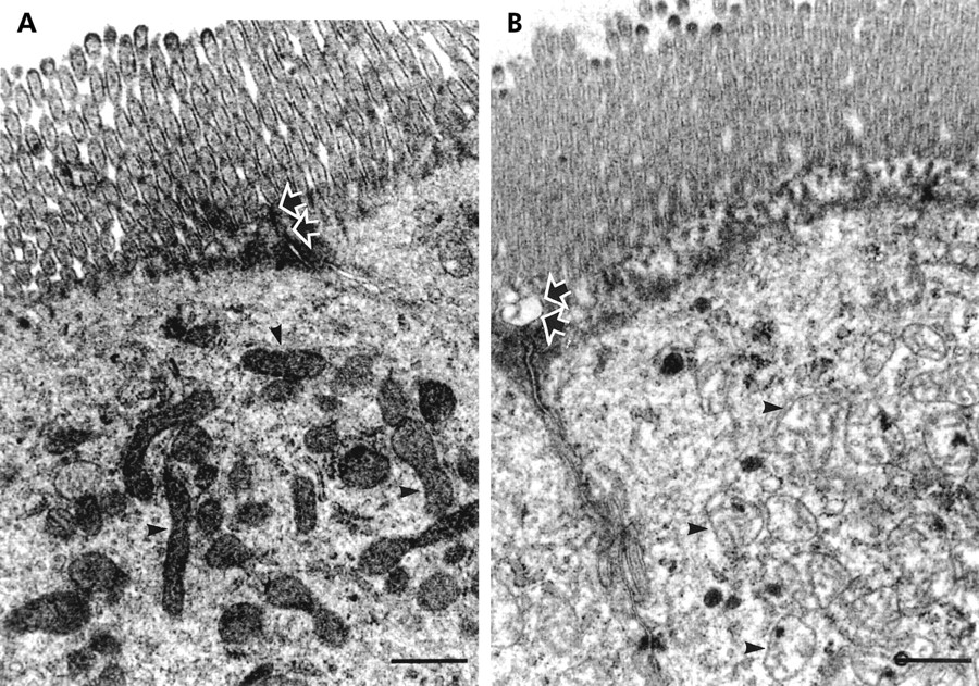
tight junction Dr.Jastrow's electron microscopic atlas

The Cell: The Histology Guide

Histology Laboratory Manual

Tight Junction - an overview | ScienceDirect Topics

Epithelial cell Zonula adherens, TEM - Stock Image - C036/7393 - Science  Photo Library
Epithelial cell Zonula adherens, TEM - Stock Image - C036/7393 - Science Photo Library

Augmented increase in tight junction permeability by luminal stimuli in the  non-inflamed ileum of Crohn's disease | Gut
Augmented increase in tight junction permeability by luminal stimuli in the non-inflamed ileum of Crohn's disease | Gut

Figure 1.2 from Role of extracellular signal-regulated kinase (ERK) in  regulation of intestinal epithelial tight junctions | Semantic Scholar
Figure 1.2 from Role of extracellular signal-regulated kinase (ERK) in regulation of intestinal epithelial tight junctions | Semantic Scholar
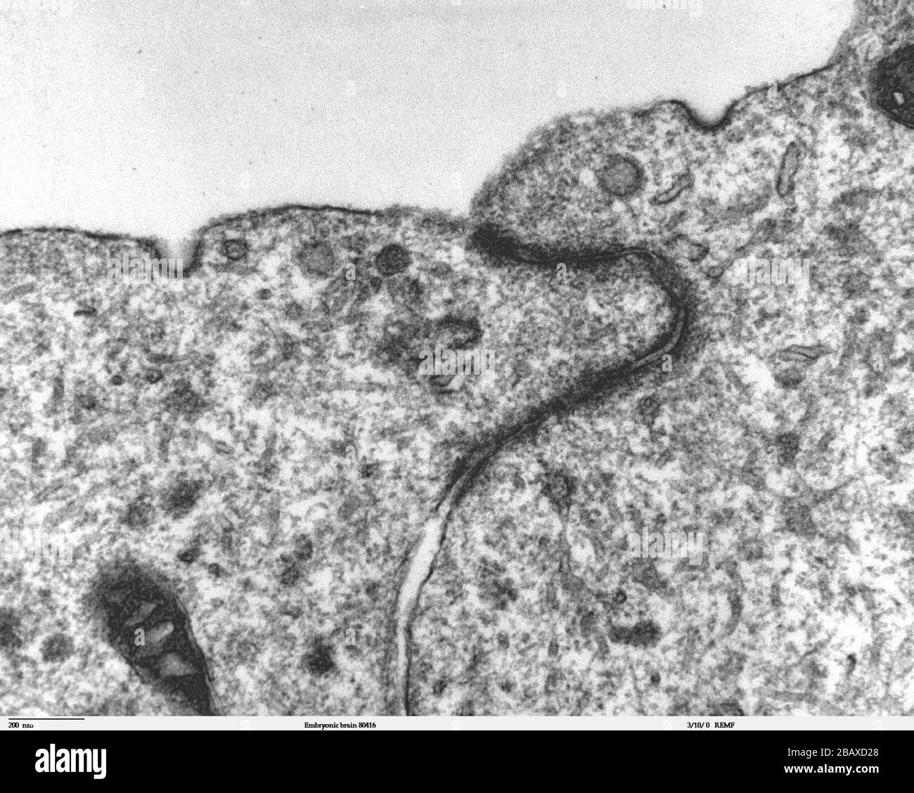
Transmission electron microscope image of a thin section cut through the  developing brain tissue (telencephalic hemisphere) of an 11.5 day mouse  embryo. This higher magnification image of Embryonic brain 80415, shows an
Transmission electron microscope image of a thin section cut through the developing brain tissue (telencephalic hemisphere) of an 11.5 day mouse embryo. This higher magnification image of Embryonic brain 80415, shows an

tight junction Dr.Jastrow's electron microscopic atlas

Electron microscopy of tight junctions of the small intestine.... |  Download Scientific Diagram
Electron microscopy of tight junctions of the small intestine.... | Download Scientific Diagram

Electron microscopy of puncta adhaerentia-type adherens junctions (AJs)...  | Download Scientific Diagram
Electron microscopy of puncta adhaerentia-type adherens junctions (AJs)... | Download Scientific Diagram

Molecular components of the adherens junction - ScienceDirect